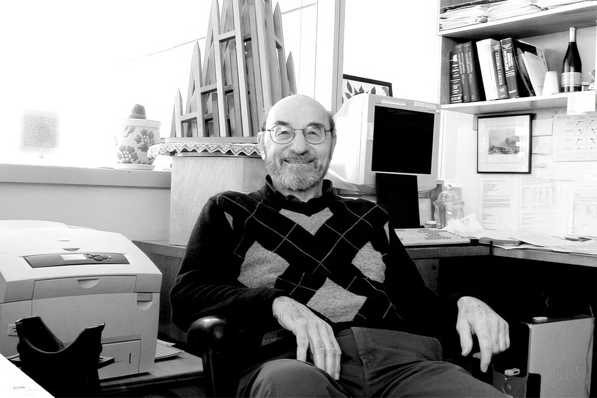
His wide-ranging and influential career included fundamental discoveries about how visual scenes and stimuli are processed from the retina through the cortical visual system.
Department of Brain and Cognitive Sciences
Peter Schiller, professor emeritus in the Department of Brain and Cognitive Sciences and a member of the MIT faculty since 1964, died on Dec. 23, 2023. He was 92.
Born in Berlin to Hungarian parents in 1931, Schiller and his family returned to Budapest in 1934, where they endured World War II; in 1947 he moved to the United States with his father and stepmother. Schiller attended college at Duke University, where he was on the soccer and tennis teams and received his bachelor’s degree in 1955. He then went on to earn his PhD with Morton Weiner at Clark University, where he studied cortical involvement in visual masking. In 1962, he came to what was then the Department of Psychology at MIT for postdoctoral research. Schiller was appointed an assistant professor in 1964 and full professor in 1971. He was appointed to the Dorothy Poitras Chair for Medical Physiology in 1986 and retired in 2013.
“Peter Schiller was a towering figure in the field of visual neurophysiology,” says Mriganka Sur, the Newton Professor of Neuroscience. “He was one of the pioneers of experimental studies in nonhuman primates, and his laboratory, together with those of Emilio Bizzi and Ann Graybiel, established MIT as a leading center of research in brain mechanisms of visual and motor function.”
Recalls John Maunsell, the Albert D. Lasker Distinguished Service Professor of Neurobiology at the University of Chicago, who did postdoctoral research with Schiller, “Peter was the boldest experimentalist I’ve ever known. Once he engaged with a question, he was unintimidated by how exacting, intricate, or extensive the required experiments might be. Over the years he produced an impressive range of results that others viewed as beyond reach.”
Schiller’s former PhD student Michael Stryker, the W.F. Ganong Professor of Physiology at the University of California at San Francisco, writes, “Schiller was merciless in his criticism of weakly supported conclusions, whether by students or by major figures in the field. He demanded good data, real measurements, no matter how hard they were to make.”
Schiller’s research spanned multiple areas. As a graduate student, he designed an apparatus, the five-field tachitoscope, that rigorously controlled the timing and sequence of images shown to each eye in order to study visual masking and the generation of optical illusions. With it, Schiller demonstrated that several well-known optical illusions are generated in the cortex of the brain rather than by processes in the peripheral visual system.
Seeking postdoctoral research, he turned to his father’s friend, Hans-Lukas Teuber, who had just accepted an offer to be founding head of the Department of Psychology at MIT. Schiller learned how to make single-unit electrophysiological recordings from the brains of awake animals, which added a new dimension to his studies of the circuitry and mechanisms of cortical processing in the visual system. Among other findings, he saw that brightness masking in the visual system was caused by interactions among retinal neurons, in contrast to the cortical mechanism of illusions.
In 1964, Schiller was appointed assistant professor. Soon after, he embarked on productive collaborations with Emilio Bizzi, who had just arrived in the Department of Psychology. Schiller and Bizzi, who is now an Institute Professor Emeritus, shared an interest in the neural control of movement; they set to work on the oculomotor system and how it guides saccades, the rapid eye movements that center objects of interest in the visual field. They quantified the firing patterns of motor neurons that generate saccadic eye movements; paired with studies of the superior colliculus, the brain center that guides saccades in primates, and the frontal eye fields of the cortex, they outlined a fundamental scheme for the control of saccades, in which one system identifies targets in the visual scene and another generates eye movements to direct the gaze toward the target.
Continuing his dissection of visual circuitry, Schiller and his colleagues traced the connections that two different types of retinal cells, known as parasol cells and midget cells, send from the retina to the lateral geniculate nucleus of the thalamus. They discovered that each cell type connects to a different area, and that this physical segregation reflects a functional difference: Midget cells process color and fine texture while parasol cells carry motion and depth information. He then turned to the ON and OFF channels of the visual system — channels originating in different types of retinal neurons: some which respond to the onset of light, others that respond to the offset of light, and others that respond to both on and off. Building on earlier work by others, and inspired by recent discoveries of ways to pharmacologically isolate ON and OFF systems, Schiller and several of his students extended the previous studies to primates and developed an explanation for the evolutionary benefit of what seems at first like a paradoxical system: that the ON/OFF system allows animals to perceive both increments and decrements in contrast and brightness more rapidly, a beneficial attribute if those shifts, for instance, represent the approach of a predator.
At the same time, the Schiller lab delved further into the role of various parts of the cortex in visual processing, especially the areas known as V4 and MT, later steps in visual processing pathways. Through single-neuron recordings and by making lesions in specific areas of the brain in the animals they studied, they revealed that area V4 has a major role in the selection of visual targets that are smaller or have lower contrast compared to other stimuli in a scene, an ability that, for example, helps an animal unmask a camouflaged predator or prey. Strikingly, he showed that many variations in images that are important for perception have a delayed influence on the responses of neurons in the primary visual cortex, indicating that they are produced by feedback from higher stages of visual processing.
Schiller’s many significant contributions to vision science were recognized with his election to the National Academy of Sciences and the American Academy of Arts and Sciences in 2007, and, in his home country, he was made an honorary member of the Magyar Tudományos Akadémia, the Hungarian Academy of Sciences, in 2008.
Schiller’s legacy is also evident in his students and trainees. Schiller counted more than 50 students and postdocs who passed through his lab in its 50 years. Four of his trainees have since been elected to the National Academy of Sciences: graduate students Larry Squire and Stryker, and postdocs Maunsell and Nikos Logothetis.
His mentorship also extended to faculty colleagues, recalls Picower professor of neuroscience Earl Miller: “He generously took me under his wing when I began at MIT, offering invaluable advice that steered me in the right direction. I will forever be grateful to him. His mentorship style was not coddling. It was direct and frank, just like Peter always was. I remember early in my nascent career when I was rattled by finding myself in a scientific disagreement with a senior investigator. Peter calmed me down, in his way. He said, ‘Don’t worry, controversy is great for a career.’ But he quickly added, ‘As long as you are right; otherwise, well …’
Schiller’s creative streak did not just influence his scientific thinking; he was an accomplished guitar and piano player, and he loved building complex and abstract sculptures, many of them constructed from angular pieces of colored glass. He is survived by his three children, David, Kyle, and Sarah, and five grandchildren. His wife, Ann Howell, died in 1999.
